
In modern dental medicine, the diagnosis and complex treatment of diseases of the face and jaws, and surgery itself, are unthinkable without the most up-to-date techniques and technology.
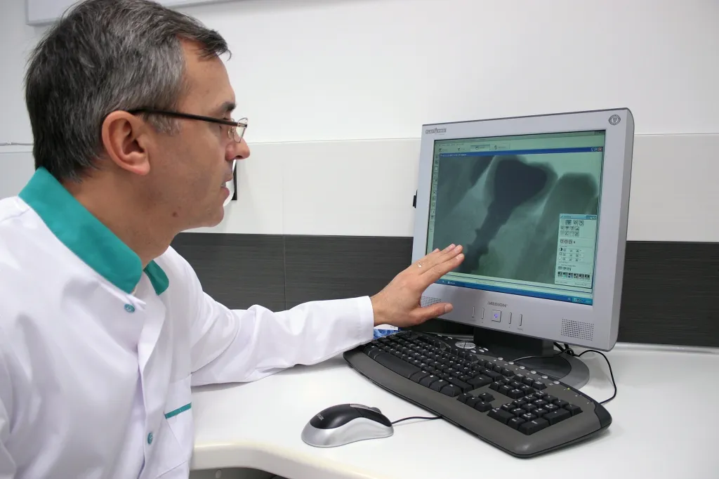
Digital X-ray unit with an intraoral sensor
With this unit our practice gets a high-quality image on a monitor with a minimal radiation dose. It helps us place the implant well in relation to the neighbouring teeth and cavities, and follow the healing after the implant is placed.
Panoramic X-ray of the jaws (orthopantomography)
Before any surgery, the position of the mandibular canal, the sinuses and in general the volume of bone in the jaws must be known. That is why a panoramic X-ray is compulsory, and sometimes it is all that implant treatment needs.
Computed tomography
This kind of X-ray uses a computer to produce detailed images of slices of the jaw (every 2 mm). The many slices give the fullest and most accurate information on the state of the alveolar bone:
- the height and width of the alveolar bone;
- the position of important anatomical structures: the mandibular canal, foramina, sinuses;
- differences in the X-ray density of tissues as small as 0.5 % can be told apart;
- computer contrast enhancement makes even very small structures visible.
A modern X-ray laboratory near our practice will do the examinations you need at short notice.